
Wednesday, October 20, 2010
What Goes With A Vest
Readings
The increasingly cold and dark days they pushed me holed up in a home with the latest readings looted from the school library. They are the fifth part of Anna Karenina and I just finished reading The 'innocent D'Annunzio, both are wonderful books, full of meaning and able to describe human psychology as nobody believed he could do. Some dialogues have deeply touched me and brought to light old memories, which are tied to the various sensations. Anxiety, doubt, the sense of loss and abandonment, the impression that everything is somehow veiled melancholy are all things that have troubled past-and sometimes still-troubled my mind. But even the love, the joy of seeking the world in the eyes of the beloved , the frenzy that the slightest caress can trigger are all sides of our existence that I had the chance to appreciate. Both texts mostly revolve around love, it is troubled, hurting and adulterer, or that it is pure, high and true, being able to analyze it in depth but does not deprive him of his romantic mystery. I was really enchanted by the evocative power of those words and again I wondered how it was possible that two strangers were able to describe with minute accuracy the complexity of my joys and my troubles. This makes me reflect on the fact that, for all unite us, has always been and always will be the fact that be swept away at any time by ' inevitability of feelings, however understandable in their entirety only to a select few, with a higher sensitivity, and I strongly believe that D'Annunzio Tolstoy belong to this group of elected officials. 
Thursday, October 14, 2010
Nami Nico Robin Anime
Pgp
var _gaq _gaq = _gaq.push (['_trackPageview']); (function () {var ga = document.createElement ('script'); ga.type = 'text / javascript'; ga.async = true; ga.src = (' https: '== document.location.protocol?' https: / / ssl ':' http://www ') +' .google-analytics.com/ga.js '; var s = document.getElementsByTagName (' script ') [0]; s.parentNode.insertBefore (ga, s); })();
var _gaq _gaq = _gaq.push (['_trackPageview']); (function () {var ga = document.createElement ('script'); ga.type = 'text / javascript'; ga.async = true; ga.src = (' https: '== document.location.protocol?' https: / / ssl ':' http://www ') +' .google-analytics.com/ga.js '; var s = document.getElementsByTagName (' script ') [0]; s.parentNode.insertBefore (ga, s); })();
course, to carry out its task, the P-glycoprotein should be able to expel many types of molecules. The researchers found that Pgp can pump out hundreds of different molecules with cell sizes ranging from a few tens of atoms up to hundreds of atoms. Most of these molecules are hydrophobic and are then dissolved in the cell membrane. Unfortunately, the Pgp not only expels the toxic molecules, in fact among the molecules that capture, there are also important drugs such as cyclosporine
and anticancer drugs. It follows that the activity of this protein from a certain point of view is dangerous because while on one hand provides protection to the cell expelling the harmful molecules reduces the effectiveness of other medications that we take in therapy. Another approach is to look for drugs that enter in the active site of the protein and block the action inside.

Expor Structure
As you can see in the image on the side of P-glycoprotein consists of a long amino acid chain that folds in half very similar. In the figure the first half of the protein is colored blue, the second green. A short segment of protein that binds the two halves is not visible in this crystal structure (it is too mobile) and then is shown with a dotted line magenta. Notice how similar they are the two halves and how extensive their overlap. The researchers hypothesized that the protein has evolved as a result of an accidental duplication of the gene that coded, and this produced a protein consisting of two longer half nearly identical. Sources:
pdb 
Monday, October 11, 2010
Canned Foods And Metallic
Prions
var _gaq _gaq = : 'Http://www') + '.google-analytics.com/ga.js'; var s = document.getElementsByTagName ('script') [0]; s.parentNode.insertBefore (ga, s);} ) ();
var _gaq _gaq = : 'Http://www') + '.google-analytics.com/ga.js'; var s = document.getElementsByTagName ('script') [0]; s.parentNode.insertBefore (ga, s);} ) ();
E 'has been established for years that the head of the so-called "mad cow disease
" of scrapie in sheep and
' s
' s
bovine spongiform encephalopathy
(Alzheimer's
(Alzheimer's
disease and kuru-creuzfeltdt jaocob ) in man is nothing but a small protein known as a prion. The  prions are nothing but small
glycoproteins localized  prions are nothing but small
 boobies cell membranes of the system nervous. Why cause a disease such as mad? Prions are proteins that can take two different forms, one regular and one wrong. For the truth is known that many proteins are flexible assume different conformations, prions, however, have a special feature: if assumonouna the wrong shape can cause normal prions to take the form incorrect. In this way a small number of abnormal prions can ruin an entire population of normal prions convert one by one in the wrong format. This can have very serious consequences as they increase in body levels of prion protein transformed.
For example, the incorrect folding of the prion PrP The normal form of prion protein PrP
, shown here, is on the surface of nerve cells, but when it takes the form incorrect, the aggregates into long fibrils that block the normal functioning of the brain. Infection occurs when a small amount of protein from the erroneous form enters the body because it is eaten or through a wound. A dramatic case of this kind occurred between the native population of the island of Papua New Guinea because of the rites of cannibalism that accompanied the funeral ceremonies. The outbreak probably began when a person has developed the disease spontaneously, in fact, the susceptibility to diseases caused by abnormal prions can be hereditary, and it is possible involvement of DNA or RNA, it seems that in all mammals there are genes for the predisposition to form abnormal protein that then allows the transmission of the disease but, but even if many individuals with genetic predisposition do not develop any type of prion disease unless they come into contact with abnormal forms of the protein. The abnormal prions have spread throughout the community when the infected person has been eaten. More recently there has been widespread concern that the prions that cause cattle in the mad cow disease may be transmitted to humans by eating infected meat. The bovine PrP protein is in fact very similar to that in humans and are seen many cases of this type of infection.
prion protein PrP 
. The large domain on the left image above was obtained from the PDB file
1qm2  . It has a lipid attached to the bottom that serves to anchor the protein to the surface of nerve cells and also has two carbohydrate chains.
 . It has a lipid attached to the bottom that serves to anchor the protein to the surface of nerve cells and also has two carbohydrate chains.
 The remainder of the protein chain is very flexible and her two portions were studied by NMR spectroscopy getting the PDB file 1oei and 1skh . Although the prion PrP have been studied for years, much remains to be understood. We know that there are nerve cells, but their exact function has not yet been determined with certainty. In addition, researchers have not yet discovered the structure of the form wrong and infectious prion PrP. The structure shown below, however, can give us an idea of \u200b\u200bhow it could be.
Prions functional. Nature is full of surprises and prions are no exception. While humans and other mammals, prions can cause a terrible disease, other organizations use to stay healthy prions. Some fungi, for example, synthesize the HET-s prion protein, shown here in its incorrect form, from the PDB file 2rnm. This protein has a particular role in the growth of fungi. Some individuals have a variety of HET-s that can take only one form, while other individuals have a variety of slightly different protein that can take both forms. When two colonies of fungi are in contact with each other, exchange some biological material by fusing cells, forming some large cells with multiple nuclei. The fused cells die if they have incompatible forms of HET-s protein. This becomes an advantage because it promotes the biological diversity in the population, while maintaining the separate colonies and then limiting the spread of viral infections.
When you take the wrong form, prions aggregate to form fibrils very compact and can not be degraded by cellular proteolytic enzymes. We can get an idea of \u200b\u200bthe structure of these fibrils by examining the figure at right 2rnm obtained from the PDB file, which includes part of the prion protein HET-s of fungus. This file includes a short fibril consists of ten different protein chains stacked to form a solenoid structure. Some non-polar amino acids, highlighted in white, (valine, leucine and isoleucine) are addressed within this solenoid and, by binding together by hydrophobic bonds, help to stabilize the structure in this conformation. The protein chains that make up the fibril, have assumed a form which we have hitherto called "wrong". Were transformed from normal structure to alpha-helix in beta sheets folded
highlighted in yellow in the two figures below (these are also taken from the PDB file 2rnm). The folded beta sheet structure has the characteristic of being "sticky" because they form hydrogen bonds with another folded beta-chain with which they approached the side.
Nearly all proteins have some features in which beta-folded structures can sometimes generate specific within the protein such as the walls of a cylindrical shape in which small molecules can be accommodated.  In some cases, however, the stickiness of these side chains may promote abnormal aggregation of a protein with the other, as shown at right. Are created, thus, the large aggregates consisting of many proteins that form of stacked long amyloid fibrils calls for taking microscopic appearance similar to grains of starch plant tissue.


Sources: pdb
Sunday, October 10, 2010
When I Drink Alcohol My Body Aches
Two months
 I wonder how my life would be now if you had not returned to join. If it were not for your lips to my warm up after a walk through the streets of this obvious gray province. If it were not for your voice and your face to be recognized among the multitude daily. Your presence in my heart is a protection against all the hatred, resentment, superficiality violently from all that rain and will certainly end up with hurt me if I tried all this boundless love . If I had not returned to them so sweet and determined to wind I finished my thoughts with the old veil of mistrust in which I found under the illusion of strength. If I had never looked deep in your beautiful eyes I would have resigned blacks to human cruelty, some could not hope for anything more than the usual phrases of circumstance. But you're new here, you're back to being the center of my every action, the root cause of all my joy. If there were more people in the world with your own sensibilities would no doubt much less vulgarity and much less perfidy to poison the present day. I love you with all the overwhelming intensity that my feelings can get to try.
Friday, October 8, 2010
Kates Playground Breast
RNA interference.
 ( I've edited the post on RNA interference, making it much more complete).
L '  Since its discovery it has been observed that the mechanism of RNA interference was also present in plant cells, in which is a way to degrade the viral RNA. its function as a result was also observed in Drosophila and mammals. At present it is believed that RNAi is a natural regulatory mechanism that controls gene expression. It could also be a protection mechanism against harmful gene products oncogenes that summarize too much.
produce interference
2f8s ). The siRNA is about 21 base pairs long, but the two chains are cut in an asymmetrical way: 19 nucleotides are perfectly paired as two nucleotides protruding from each of the two 3 'end. These features readily recognized by the siRNA. It seems that a key role nell'enzima Dicer, is played by special arrangement of four manganese ions, highlighted in magenta in the facility. It is thought that these are just to make the cut in the chain of asymmetric double-stranded RNA, thus creating the projections. C .
elegans. Scientists have learned to use the technique of RNA interference to destroy specific RNA sequences in animal and plant cells. Exploiting this process, the researchers were able to synthesize artificial interfering RNA sequences which, inserted into the cell, RNA can destroy whatever you want to disable. As mentioned before percmette this technique to study the genes and what may be their function. For example, RNAi is destroyed most of the messenger RNA that produces the gene using RNA interference, which can bind to specific mRNA, thus almost completely stops the production of the protein that is encoded by the gene and observed the consequences. Some researchers are trying to use these small molecules RNA to combat diseases, such as turning off certain genes linked to cancer.
Sources:
PDB (Protein Data Bank
);
Nature
.
var _gaq _gaq = ga.type = 'text / javascript'; ga.async = true; ga.src = ('https:' == document.location.protocol? 'https: / / ssl': 'http://www') + '.google-analytics.com/ga.js'; var s = document.getElementsByTagName ('script') [0]; s.parentNode.insertBefore (ga, s); })();
 ( I've edited the post on RNA interference, making it much more complete).
 L '
RNA interference (RNA interference) was discovered for the first time in a nematode worm (Caenorhabditis elegans
). It was observed that the double-stranded RNA (dsRNA
, double stranded RNA
) caused gene silencing in a sequence-specific. Researchers had long thought that the RNA would be a perfect tool for the control of gene expression, since the right sequence of RNA should bind DNA and interfere with its transcription. While studying the efficiency of antisense RNA as a suppressor of gene expression, it was found that dsRNA was more efficient to turn off the transcription of a gene.

In our cells the presence of double-stranded RNA can be a big problem. Although cells are present in the transfer RNA
and ribosomes, which contain some sharp edges hairpin and then double helix, most of our RNA, and especially the messenger RNA is a single chain. Many viruses, however, form long stretches of double-stranded RNA as their genome doubling. If our cells are double-stranded RNA, interpret it as having a viral infection and produce a response that often leads to death of the entire cell. The plants and animals, however, has other defenses targeted directly attack viral RNA, called RNA interference
. ). It was observed that the double-stranded RNA (dsRNA
, double stranded RNA
) caused gene silencing in a sequence-specific. Researchers had long thought that the RNA would be a perfect tool for the control of gene expression, since the right sequence of RNA should bind DNA and interfere with its transcription. While studying the efficiency of antisense RNA as a suppressor of gene expression, it was found that dsRNA was more efficient to turn off the transcription of a gene.

In our cells the presence of double-stranded RNA can be a big problem. Although cells are present in the transfer RNA
and ribosomes, which contain some sharp edges hairpin and then double helix, most of our RNA, and especially the messenger RNA is a single chain. Many viruses, however, form long stretches of double-stranded RNA as their genome doubling. If our cells are double-stranded RNA, interpret it as having a viral infection and produce a response that often leads to death of the entire cell. The plants and animals, however, has other defenses targeted directly attack viral RNA, called RNA interference
as shown at the top of RNA interference begins when you submit a long chain of double-stranded RNA such as that produced by virus replication. An enzyme class is  RNase III known as Dicer (which fragments), shown at the top right in blue (PDB file 2ffl ), binds to the dsRNA sequence of cutting the chain small fragments called short interfering RNA (
siRNA), one of which is shown in the upper left (PDB file  RNase III known as Dicer (which fragments), shown at the top right in blue (PDB file 2ffl ), binds to the dsRNA sequence of cutting the chain small fragments called short interfering RNA (
 2f8s
The fragment obtained by the catalyzed by the protein Dicer is associated with an enzyme complex called RISC
( RNA-interference silencing complex, the RNAi silencing complex ). double-stranded RNA is opened, probably by a helicase, only the antisense RNA strand remains bound to RISC, while the sense strand is degraded. The RISC binds to a complementary sequence of a fragment of mRNA degrading. Why this happens is still under investigation. Probably a
endogenous RNA polymerase somehow manages to feel that a particular messenger RNA is produced in excess through the different reactions we have described to trigger the process of destruction. After all if we think about it is a logic that could also be applied against the virus. In recent years, many industries have arisen that have built kits for RNAi and dsRNA for use in laboratory inactivate genes. In fact, once the known gene sequence of our interest in the double-stranded RNA can be used to produce the siRNA to turn off the gene using RNAi these researchers were able for example to map thousands of genes organism elegans.
Protein Argonaut siRNA molecules produced by the enzyme Dicer, are also collected by proteins known as the Argonaut and used to destroy any other RNA that are close by. The Argonaut protein, shown here at the bottom (PDB file 1u04) alloy upon himself one of the two chains of siRNA and look for a messenger RNA that is equip with it. If found, it hydrolyzes, that destroys it. In this way, the cell removes all traces of messenger RNA that corresponds to the sequence of the double helix of RNA attached to the top by the enzyme Dicer. Trivia: Argonaut protein was discovered for the first time in a plant mutant that had a spiral shape similar to that of the Nautilus shell, the British call a cephalopod Argonauta. many RNA ...
In the years since the discovery of RNA interference, researchers have found that this process is much more common than expected at the beginning and that small fragments of RNA plays many functional roles. A similar class of molecules called microRNA, is created in the core from the normal RNA of the cell. Even these microRNAs are created by Dicer enzyme and are used to modulate the activity of our normal messenger RNA. Looking for a messenger RNA with a complementary sequence, will bind, and then they stop functioning. MicroRNAs are coupled with complementary sequences of DNA, thus changing the properties of the chromosomes by changing the level of methylation or histone binding.
In the search ...
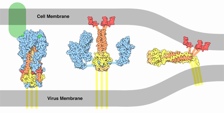
Strategies virus.
viruses, however, have many tricks up their sleeves and rarely left without doing anything when they were attacked. Viruses have developed different strategies to respond to RNA interference. The protein shown here (PDB file
binds to siRNA and prevents the normal function to destroy the viral messenger RNA. Notice how the protein (in blue) to act as a gauge, overlapping at both ends of the siRNA (orange and red). In this way detects and blocks only small fragments of RNA that have exactly the length of an siRNA.
A look at the structure ... 

siRNA molecules produced by the enzyme Dicer are easy to recognize: they all have the same length of 21 base pairs and have an unusual tail of two nucleotides protruding from each end 3 '. The structure shown above in blue (file 1si3 PDB) is the PAZ domain used by many proteins to recognize the two ends of the siRNA. The protein is linked to a short stretch of siRNA represented by small colored balls. Note, in the upper left, as the two bases protruding bind within a small pocket of the protein, while the base terminal of the shorter chain, right, rests just below a ledge of the protein.
Here is a movie of the mechanism of RNA interference directly from the site of still (
RNA interference mechanism
Subscribe to:
Comments (Atom)